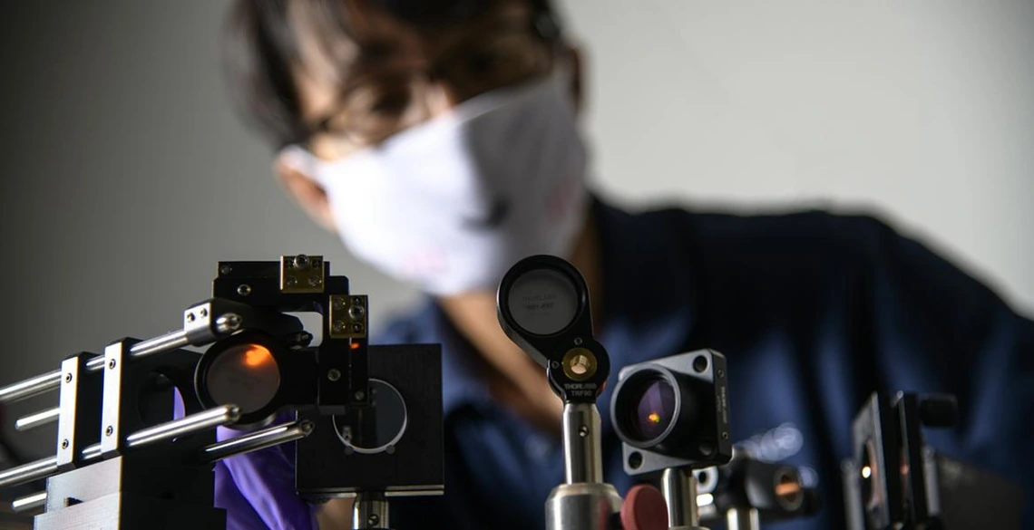
Kang Leads $1M Project to Fight Vision Loss

BME associate professor Dongkyun "DK" Kang is on a mission to diagnose and treat vision loss in a way that is faster and cheaper than currently possible. With a $1 million grant from the National Eye Institute, Kang is creating a portable device that can treat corneal ulcers, which cause blindness and visual impairment in 4.3 million people worldwide every year.
The first-line diagnostic tool for corneal ulcers is a slit lamp — a device used in routine eye exams to shine a slit of light, which the ophthalmologist can move back and forth over a patient's eye to look for problems. However, even experienced ophthalmologists often can't determine the origin of an infection from slit lamp examination alone. Knowing whether the infection is fungal, parasitic or bacterial is key to treating the ulcer effectively. Today's gold standard is microbiological testing, which involves scraping off a small piece of the cornea and culturing it for several weeks. However, Kang says neither of these methods are ideal.
Confocal microscopes in use now achieve high resolution and contrast by illuminating just one point at a time with a laser-focused light. The laser moves over each point to gather information, combines all the data, and digitizes it to create a highly detailed image. This high level of resolution is critical, but the required mechanical precision is time-consuming. Physicians can only capture a relatively small area in the image. Eye movements, which are practically inevitable when a device is touching a patient's eye, often result in physicians having to rescan some areas, making the process even longer. And it's costly. Confocal microscopes run upwards of $75,000, severely limiting their use in rural clinics.
The key to the technology Kang is working on is that it scans in two dimensions rather than one – line by line, rather than dot by dot. The "portable in vivo confocal ophthalmoscope" device uses a diffraction grating to disperse the light source into different colored wavelengths, similar to what happens when light passes through a prism. The diffraction illuminates a two-dimensional line, rather than a one-dimensional point, so a much larger portion of the eye can be scanned at a time without sacrificing image quality.
"Maybe 10 or 20 years down the road, each eye doctor will have one of our devices and take a look at patients' cornea in a very high resolution," Kang said in an interview with KGUN9. "That can really help patients in terms of diagnosis and treatment."
