
BME Researchers Detect Urethral Injury with Innovative Imaging Technique
BME researchers used optical coherence tomography (OCT) to identify urethral electrothermal injury.
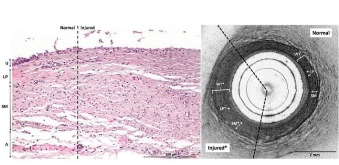
Urethral electrothermal injury shown through optical coherence tomography.
In a new study, researchers from BME have explored a novel approach to detect electrothermal ureteral injuries, a common complication during pelvic surgery.
The ureters, delicate tubes that transport urine from the kidneys to the bladder, are particularly vulnerable due to their proximity to other anatomical structures. Unfortunately, current detection methods often fall short in promptly identifying subtle thermal injuries, which can take days or even weeks to manifest.
Enter optical coherence tomography (OCT) endoscopy—a minimally invasive imaging technique that may revolutionize ureteral injury detection. In research reported in Biophotonics Discovery (BIOS), an interdisciplinary team led by researchers from BME externally applied electrothermal energy to explanted pig ureters, simulating a spectrum of injury severity. Immediately afterward, they performed OCT endoscopy and compared the resulting images to histology as the gold standard for interpretation.
The findings were remarkable. Lesion size on OCT images correlated with the treatment power used, providing a quantitative measure of injury severity. Moreover, qualitative markers of injury were detectable in nearly all cases, except for one lesion treated at low power.
Dig into BME's novel research here.